(A01-1)
Stroke, myocardial infarction, and cancer are the three major diseases associated with adult lifestyle habits. Final diagnoses for many diseases, including these three, are usually made with microscopic high-resolution imaging of the associated lesions. Nevertheless, it is often very difficult to obtain histological samples because of the invasiveness of the procedure needed to obtain a specimen. Therefore, there is a real need for noninvasive diagnostic imaging providing microscopic resolution. Magnetic resonance imaging (MRI) is one of the essential imaging techniques in modern medicine and has been used to visualize a wide variety of biological phenomena noninvasively. Its temporal and spatial resolution, however, are not sufficient for the observation of cytopathological changes.
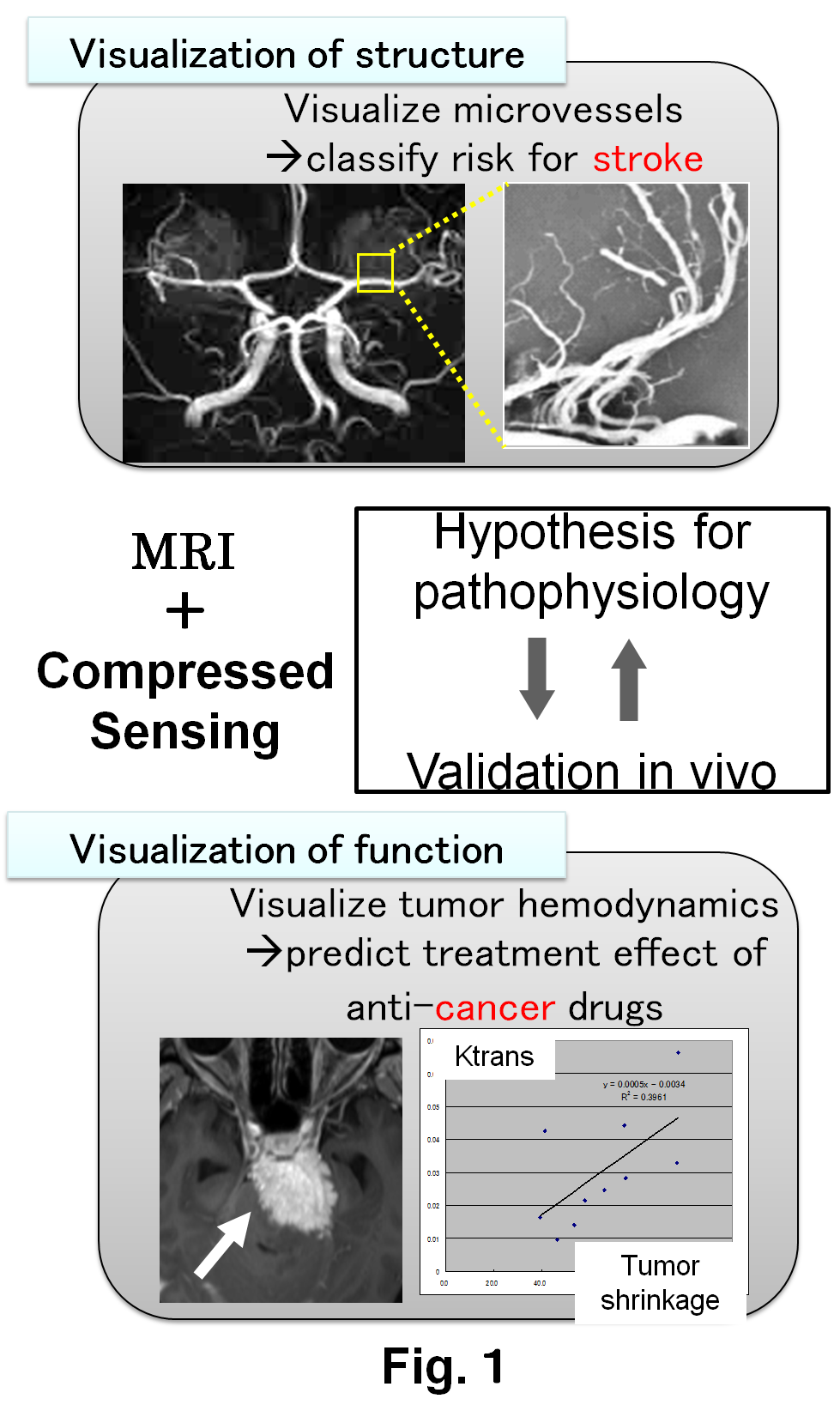
By taking advantages of compressed sensing, which is a recently developed technique for data processing, we aim to increase the spatial and temporal resolution of MRI so that MRI can be used more effectively in the noninvasive visualization of structure and function of the diseases, in the diagnosis and early treatment of diseases, and in preventive health-care.
Abundant hydrogen nuclei exist in vivo. By manipulating these hydrogen nuclei with radiofrequency (RF) microwaves under a static magnetic field, MRI collects signals from the human body. Cross-sectional images are obtained by applying Fourier transformation to the collected data. Recently, compressed sensing is attracting attention because it can dramatically reduce MRI scan time (Lustig et al, MRM 2007). This technique enables reconstruction of the MR images from under-sampled data with fewer artifacts, by making use of sparsifying transforms such as Wavelets.
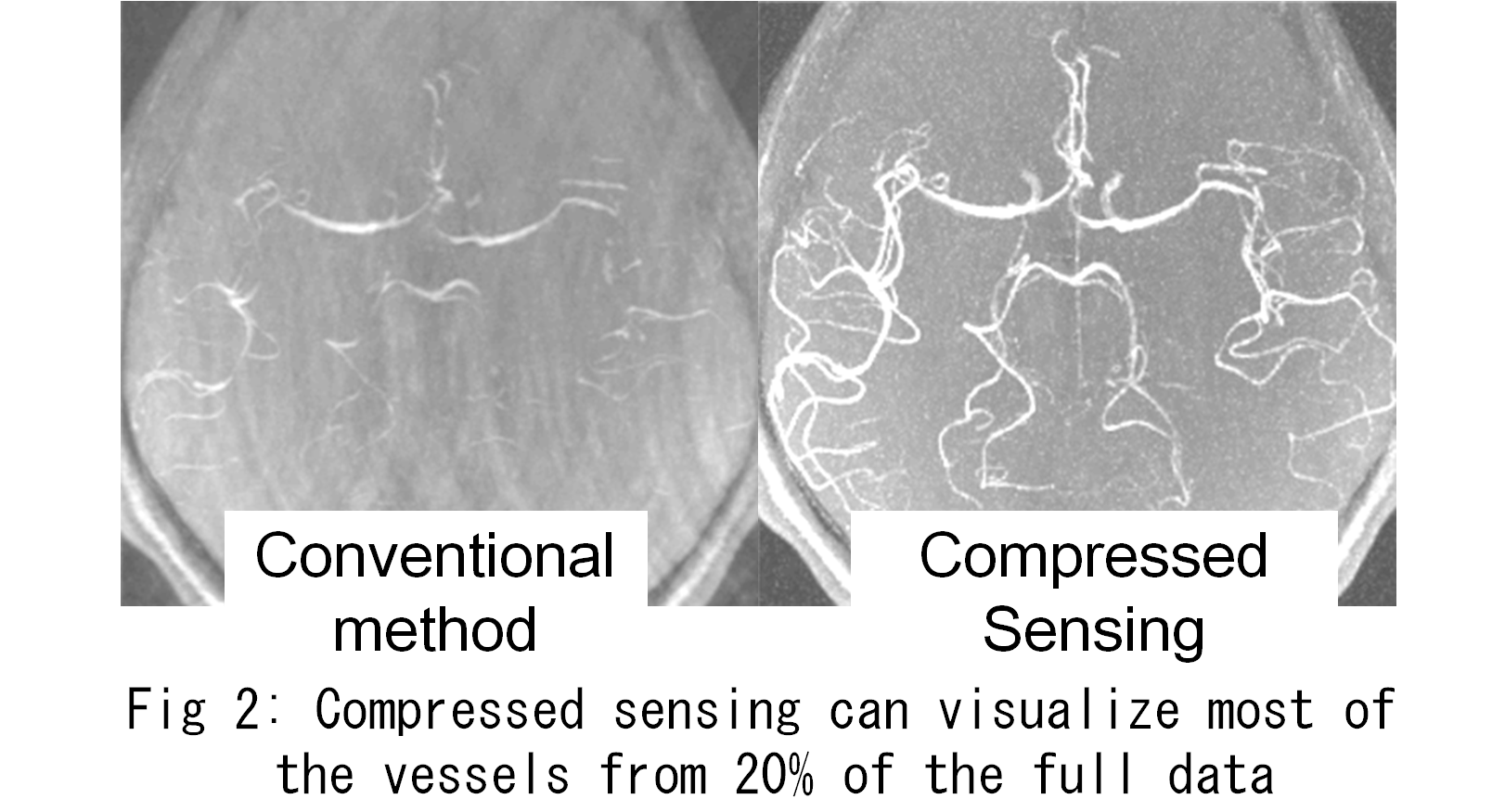
We have reported decreased visualization of lenticulostriate arteries in subjects with hypertension. To achieve more detailed visualization of lenticulostriate arteries, we started basic experiments with professor Tanaka, the of the (B01-1). Preliminary results are shown in Figure 2.
Through this initial experience, we have noticed that previously reported method to minimize residuals in compressed sensing does not yield visualization of microvessels of our interest. By combining the viewpoints of medical doctors and informatics scientists, we should be able to visualize fine structures of lesions (e.g., microvessels) and organ-specific characteristics (e.g., tissue perfusion). Promoting this project, not only in medical imaging but also in other natural sciences in collaboration with other teams, would cultivate a new area of science.
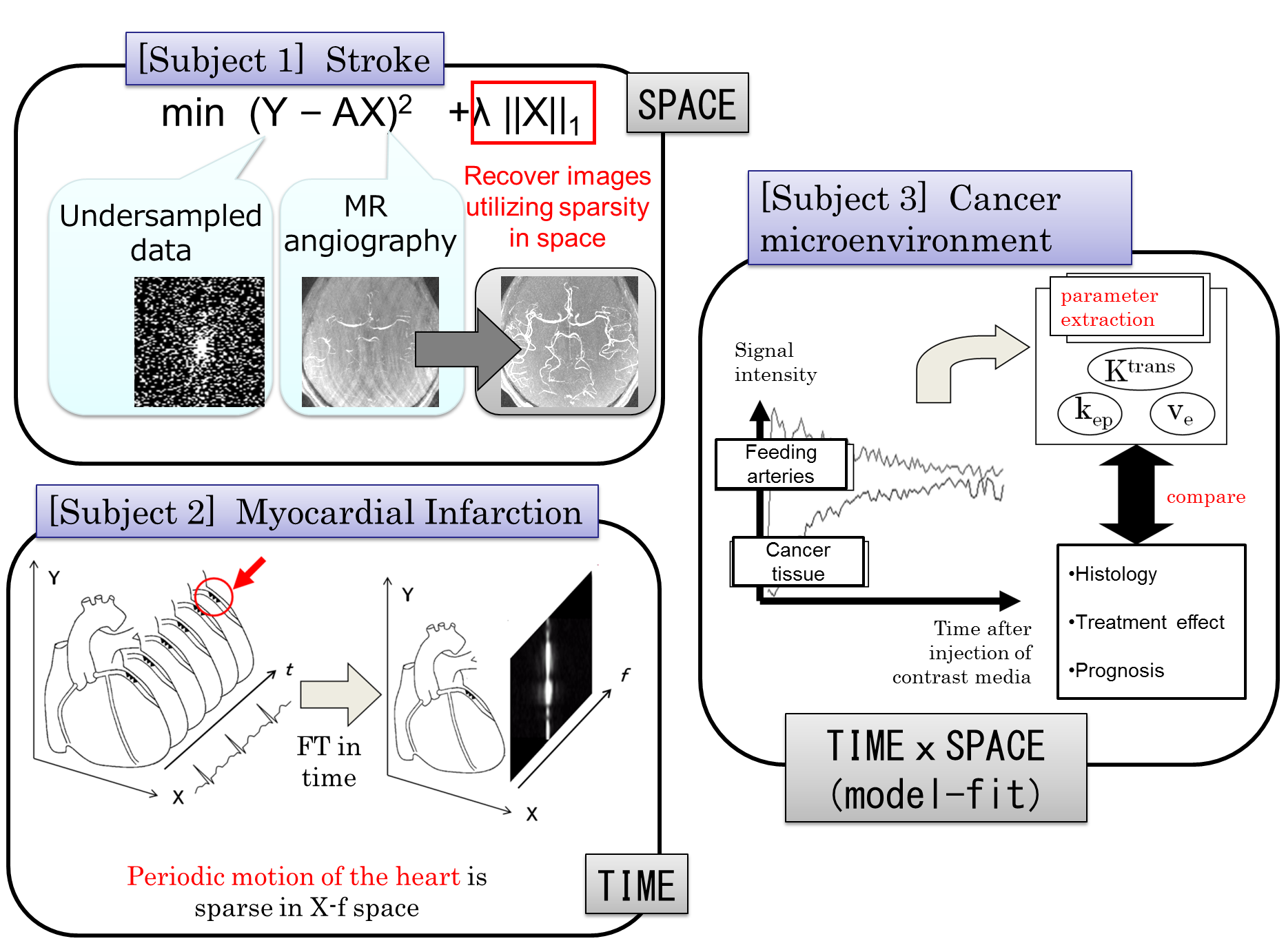
Abnormality in vessels are associated with three major adult diseases (stroke, myocardial infarction, and cancer), but mechanisms of pathophysiology differ in these diseases. By focusing on sparsity of vessels in time and space, we have identified clinical applications and corresponding technical challenges that need to be solved.
- [Subject 1] Stroke: High-resolution and high-speed three-dimensional imaging of cerebral vessels by utilizing spatial sparsity
We will focus on the spatial sparsity of cerebral vessels. Visualization of clinically important microvessels is investigated. Methods to accelerate high-resolution imaging will be developed by investigating relationships between sparsity, undersampling rate, and basis function. - [Subject 2] Myocardial infarction: High-speed imaging of organs in periodic motion
We will take advantage of the periodic motion of the heart and develop methods for efficient data acquisition as well as reconstruction algorithms appropriate for temporally sparse data. - [Subject 3] Microvessels in cancer: Data acquisition for model-based data analysis
In a series of dynamic MR imaging, the signals from cancer tissue and its feeding vessels increase after the administration of contrast media. Model-based analysis such as pharmacokinetic analysis are applied to extract several parameters. By focusing on the redundancy in the acquired data, and by using the results obtained in subjects 1 and 2 as well as a method of sparse modeling, we will investigate in improving the temporal and spatial resolution of MRI.